Cell Membrane Proteins Imaged in 3D
08/13/2020
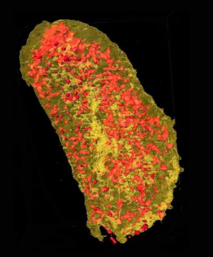
Ultrabright X-rays revealed the concentration of erbium (yellow) and zinc (red) in a single Escherichia coli cell expressing a lanthanide-binding tag and incubated with erbium. [Reprinted with permission from Victor et al. 2020. “Lanthanide-Binding Tags for 3D X-Ray Imaging of Proteins in Cells at Nanoscale Resolution,” Journal of the American Chemical Society 142(5), 2145–49. Copyright 2020 American Chemical Society. DOI:10.1021/jacs.9b11571]
The Summary
Using ultrabright X-rays, researchers at the National Synchrotron Light Source II have demonstrated a new technique for imaging proteins in three dimensions with nanoscale resolution. Their approach, which employs X-ray fluorescence microscopy, enables scientists to identify the precise location of proteins within individual cells, reaching the resolution of the cell membrane and the smallest subcellular organelles.
Watch a 3-D X-ray movie of a single E.coli cell imaged at NSLS-II with a sub-15 nm X-ray beam, which is the highest resolution X-ray fluorescence tomogram of a biological cell ever collected (as of the paper’s publication date in 2020).
Facilities and Instruments
Two-dimensional X-ray fluorescence microscopy was performed at the Bionanoprobe (BNP) at beamline 21-ID-D (now at 9-ID-B) at the Advanced Photon Source at Argonne National Laboratory. Three-dimensional X-ray nanotomography was performed at the Hard X-ray Nanoprobe beamline 3-ID at the National Synchrotron Light Source II (NSLS-II) at Brookhaven National Laboratory.
Related Links
- BER Resource: Center for BioMolecular Structure
- BER Resource: Structural Biology Center
- Feature Story: Scientists Produce 3-D Chemical Maps of Single Bacteria
- News: Cell Membrane Proteins Imaged in 3-D
References
Victor, T. W., et al. 2020. “Lanthanide-Binding Tags for 3D X-Ray Imaging of Proteins in Cells at Nanoscale Resolution,” Journal of the American Chemical Society 142(5), 2145–49. [DOI:10.1021/jacs.9b11571]