X-ray Nanofluorescence Tomography of Single Bacterial Cells
09/07/2018
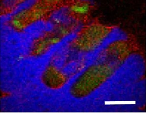
An X-ray fluorescence image showing the co-localization of zinc (green), calcium (red), and chlorine (blue) in E. coli cells. The four cells shown in the image lie parallel to each other and are embedded in sodium chloride crystals. [Reprinted under a Creative Commons Attribution 4.0 International (CC BY 4.0) license from Victor, T. W., et al. 2018. "X-ray Fluorescence Nanotomography of Single Bacteria with a Sub-15 nm Beam." Scientific Reports. 8: 13415. DOI:10.1038/s41598-018-31461-y.]
X-ray fluorescence (XRF) nanotomography was used to image elemental distribution in individual E. coli bacterial cells using a sub-15 nm beam at the Hard X-ray Nanoprobe beamline (HXN, 3-ID) at the National Synchrotron Light Source II. The measurements were simultaneously combined with ptychography to image the cells’ structural components. The results showed a generally uniform distribution of calcium but an inhomogeneous zinc distribution, most notably with concentrated regions of zinc at the polar ends of the cells.
XRF microscopy is a growing approach for imaging the trace element concentration, distribution, and speciation in biological cells at the nanoscale. Moreover, three-dimensional nanotomography provides the added advantage of imaging subcellular structure and chemical identity in three dimensions without the need for staining or sectioning of cells.
The work demonstrates that simultaneous two-dimensional ptychography and XRF nanotomography can be performed with a sub-15 nm beam size on unfrozen biological cells to co-localize elemental distribution and nanostructure simultaneously. This multimodal approach presents new possibilities in understanding subcellular biochemistry in individual organelles, which are usually analyzed at the population level.
Related Links
- BER Resource: Center for BioMolecular Structure
- Feature Story: Scientists produce 3-D chemical maps of single bacteria
- News: First X-ray nanofluorescence tomography of single bacteria
References
T. W. Victor, L. M. Easthon, M. Ge, K. H. O’Toole, R. J. Smith, X. Huang, H. Yan, K. N. Allen, Y. S. Chu, L. M. Miller. “X-ray Fluorescence Nanotomography of Single Bacteria with a Sub-15 nm Beam.” Scientific Reports. 8: 13415 (2018). [DOI: 10.1038/s41598-018-31461-y]