Multimodal X-Ray Imaging Technique for Single Cells
03/07/2024
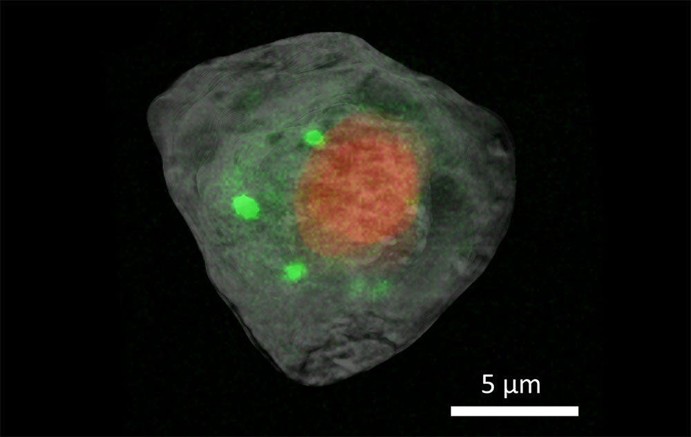
Integrated X-Ray Imaging Techniques Reveal Structural and Chemical Cellular Characteristics. Superimposed X-ray computed tomography images (gray and red coloring) and X-ray fluorescence microscopy images (green coloring) show the nucleus (red) and cytosol (gray) with calcium distribution (green) in a single human embryonic kidney cell.
[Reprinted under a Creative Commons license (CC BY) from Lin et al. 2024. DOI:10.1038/s42003-024-05950-y]
The Science
Scientists from DOE’s Brookhaven National Laboratory used X-ray computed tomography (XCT) and X-ray fluorescence (XRF) microscopy to develop a novel multimodal method to noninvasively image a single cell, achieving nanometer resolution without damaging delicate organelles. The method demonstrates the feasibility and broad applicability of using X-rays to understand cellular structures and the roles of chemical elements and related proteins in signaling and other biological processes.
The Impact
Understanding the inner workings of cells and their interactions at such detailed levels can transform the approach to various fields, particularly in identifying how pathogens interact with host cells. This multimodal imaging approach provides insights that could improve agricultural practices by studying plant–pathogen interactions and enhance drug delivery systems. These insights could lead to better strategies for improving crop resilience and combating infectious diseases.
The Summary
Cells from the human embryonic kidney (HEK) 293 line were chemically preserved using paraformaldehyde to maintain cellular structure. They were then rapidly frozen, transferred to liquid nitrogen, and freeze-dried before being placed under a microscope to locate and label them for targeted imaging. Standardized sample holders were created for use on multiple pieces of equipment to ensure cells could be reliably held in place for multiple measurements.
The team used two imaging techniques found at NSLS-II: XCT and XRF microscopy. The Full Field X-ray Imaging (FXI) beamline was used to collect XCT data. The technique uses X-rays to show a cross-section of a solid sample, providing information about a cell’s physical structure. This is similar to computed tomography scans in medical imaging.
The researchers also used the Submicron Resolution X-ray Spectroscopy (SRX) beamline to collect XRF microscopy data, which provides information about the distribution of chemical elements within a cell. XRF directs high-energy X-rays at a sample, exciting the material and causing it to emit X-ray fluorescence. This X-ray emission has a unique signature, enabling scientists to determine exactly which elements comprise the sample and how they are distributed to fulfill their biological functions.
Achieving high-resolution fluorescence maps of multiple cells can be very time consuming, even for two-dimensional images. The XCT technique, which produces three-dimensional images, can help speed the process. The information can help guide two-dimensional fluorescence measurements to specific locations of interest. This saves time, increases throughput, and reduces the length of time samples are exposed to X-rays, which mitigates potential damage to fragile cells.
Funding
This work was supported by the Laboratory Directed Research and Development Program LDRD21-013 of Brookhaven National Laboratory and the DOE Biological and Environmental Research program (BER; KP1601011). The work used the Full-field X-ray Imaging (FXI) beamline at 18-ID and the Submicron Resolution X-ray Spectroscopy (SRX) beamline at 5-ID of the National Synchrotron Light Source II, a DOE Office of Science user facility operated by Brookhaven National Laboratory under Contract No. DE-SC0012704. The work used the Vitrobot at the Laboratory for BioMolecular Structure, which is supported by BER (KP1607011). The work used the confocal microscope at the Center of Functional Nanomaterials, which is a DOE Office of Science user facility at Brookhaven National Laboratory under Contract No. DE-SC0012704.
Related Links
- BER Resource: Laboratory for BioMolecular Structure
- Pioneering the Cellular Frontier: Brookhaven Researchers Explore a Single Cell Using Advanced X-Ray Imaging Techniques
References
Lin, Z., Zhang, X., Nandi, P., et al. 2024. “Correlative Single-Cell Hard X-Ray Computed Tomography and X-Ray Fluorescence Imaging,” Communications Biology 7(280). DOI: 10.1038/s42003-024-05950-y