Revealing How Bacterial Organelles Assemble
06/23/2017
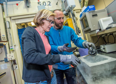
Structure of microcompartment’s protein shell could help research in bioenergy, pathogenesis, and biotechnology. Cheryl Kerfeld and Markus Sutter handle crystallized proteins at Berkeley National Laboratory’s Advanced Light Source. [Image courtesy Marilyn Chung, Lawrence Berkeley National Laboratory]
The Summary
Scientists are providing the clearest view yet of an intact bacterial microcompartment, revealing the structure and assembly of the organelle’s protein shell at atomic-level resolution. They studied the “photogenic” organelle shell of an ocean-dwelling slime bacteria Haliangium ochraceum.
Providing the first view of the shell of an intact bacterial organelle membrane, this full structural view can help provide important information for beneficial use in fighting pathogens or bioengineering bacterial organelles.
The research team said these organelles, or bacterial microcompartments (BMCs), are used by some bacteria to fix carbon dioxide. Thus, understanding how the microcompartment membrane is assembled, as well as how it lets some compounds pass through while impeding others, could contribute to research in enhancing carbon fixation and, more broadly, bioenergy.
This class of organelles also helps many types of pathogenic bacteria metabolize compounds that are not available to normal, nonpathogenic microbes, giving the pathogens a competitive advantage.
Instruments and Facilities
Michigan State University–DOE Plant Research Laboratory and the Molecular Biophysics and Integrated Bioimaging Division at Lawrence Berkeley National Laboratory; Stanford Synchrotron Radiation Lightsource.
Funding
Support: National Institutes of Health’s (NIH) National Institute of Allergy and Infectious Diseases (NIAID) grant 1R01AI114975-01 and the Office of Basic Energy Sciences (OBES), U.S. Department of Energy (DOE) Office of Science, under contract no. DE-FG02-91ER20021. Advanced Light Source (ALS), Lawrence Berkeley National Laboratory (LBNL) support: OBES, Director, DOE Office of Science, under contract no. DE-AC02-05CH11231. B.G. support: advanced postdoctoral mobility fellowship from Swiss National Science Foundation (NSF; project P300PA_160983). Use of Stanford Synchrotron Radiation Lightsource (SSRL), SLAC National Accelerator Laboratory (SLAC) support: OBES, DOE Office of Science, under contract no. DE-AC02-76SF00515. M.S. and C.A.K.: inventors on patent application 62509553 submitted by LBNL that covers strategies for scaling the shell-protein system described in this work. Cryo-EM map of complete shell deposited at Electron Microscopy Data Bank (EMDB) with accession code EMD-8747. X-ray crystallographic coordinates and structure-factor files deposited in Protein Data Bank (PDB) under the following accession numbers: 5V74 (complete shell), 5V75 (BMC-T2), and 5V76 (BMC-T3).
Related Links
- BER Resource: Structural Molecular Biology Resource
- News: Study Sheds Light on How Bacterial Organelles Assemble
References
Sutter, M., et al. “Assembly Principles and Structure of a 6.5-MDa Bacterial Microcompartment Shell.” Science 23(6344), 1293–1297 (2017). [DOI:10.1126/science.aan3289].