Soft X-ray Tomography Captures Mesoscale Organelle Interactions
04/07/2022
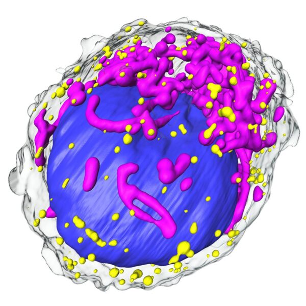
Soft X-ray tomography combines rapid collection and high-resolution visualization to reveal the shape of cellular substructures and their interactions, such as in this pancreatic beta cell (nucleus, blue; mitochondria, magenta; insulin vesicles, yellow). [Reprinted under a Creative Commons Attribution 4.0 International License (CC BY 4.0) from V. Loconte, et al. 2022. DOI: 10.1016/j.str.2022.01.006]
The Summary
Inter-organelle interactions are a vital part of normal cellular function but are difficult to quantify due to the range of scales encountered in cell biology and the limitations of traditional imaging approaches. Soft X-ray tomography (SXT) can rapidly map ultrastructural reorganization and inter-organelle interactions in intact cells by taking advantage of the naturally occurring, differential X-ray absorption of carbon-rich compounds in each organelle.
As an example, SXT was used to map the spatiotemporal evolution of insulin vesicles and their co-localization and interaction with mitochondria in pancreatic beta cells during insulin secretion and in response to different stimuli. The technique enabled quantification of changes in the morphology, biochemical composition, and relative position of mitochondria and insulin vesicles. The findings highlight the importance of comprehensive and unbiased mapping at the mesoscale to characterize cell reorganization that would be difficult to detect and quantify with other existing methodologies.
The same technique can be useful for quantifying morphological changes inside a microbial cell in response to genomic manipulations or other perturbations.
Related Links
- BER Resource: National Center for X-Ray Tomography
- Feature Story: Soft X-rays capture the dance of the organelles
References
Loconte, V. et. al. 2022. Soft X-ray tomography to map and quantify organelle interactions at the mesoscale. Structure, 30, 1–12. [DOI: 10.1016/j.str.2022.01.006]