Understanding an Essential Chaperone Complex
09/30/2020
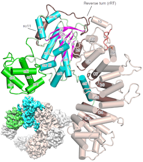
Lower left inset: cryo-EM 3D map of the Ric-8A:Gα:4Nb complex (colored wheat:cyan:green, respectively) and nanobodies colored gray. Rest of image: the annotated ribbon and cylinder drawing shows the same complex produced from X-ray diffraction data. [Reprinted from L. J. McClelland et al. 2020. DOI: 10.1038/s41467-020-14943-4 (CC BY 4.0).]
The Science
Scientists reveal a protein binding mechanism connected to embryo development and cancer. The structure of the chaperone protein Ric-8A bound to a G-protein-alpha subunit (Gα) reveals the mechanism for Gα activation through phosphorylation of Ric-8A.
The Impact
Ric-8A is a protein involved in the regulation of cell division, which is essential for embryo development. This structure reveals how it acts as a chaperone for Gα in this process.
The Summary
Many modern developments in biology, medicine, and biotechnology have been made possible through our understanding of how biological structures, such as proteins in cells, interact with each other. But to truly reveal their function and the role they play in diseases and medical conditions, scientists need to visualize these structures at the atomic level.
This is especially important for more elusive proteins and their functions, such as the Ric-8A protein, a Guanine Nucleotide exchange factor that serves as a chaperone for Gα. Gα are located at membranes and inside cells and are an essential part of several cellular signaling systems. Ric-8A is essential for asymmetric cell division in the process of embryonic development and is also a potential therapeutic target for certain cancers.
In a study, scientists revealed the structure and binding mechanism of the Ric-8A protein to the Gαi1 complex. The team used a combination of cryo-electron microscopy and x-ray studies at various light source facilities, including x-ray crystallography at the Frontier Microfocusing Macromolecular Crystallography (FMX) beamline. The FMX beamline is part of the advance life science suite of beamlines at the National Synchrotron Light Source II (NSLS-II), a U.S. Department of Energy (DOE) Office of Science User Facility located at DOE’s Brookhaven National Laboratory.
The researchers discovered that the mechanism of Ric-8A differs from the usual mechanism that the G protein uses at its receptors. Ric-8A engages a specific conformation of Gα at multiple interfaces to form a complex that is stabilized by phosphorylation within a Ric-8A segment that connects two Gα binding sites.
Funding
The FMX (17-ID-2) beamline is supported by NIH grant P41GM111244 and the Department of Energy (DOE), KP1605010. T.I.D. is supported by the SSRL Structural Molecular Biology Program by the DOE and by NIH grant P41GM103393. Small angle X-ray scattering experiments (SAXS; aka. solution x-ray scattering) were conducted at the Advanced Photon Source, operated for the DOE Office of Science by Argonne National Laboratory under Contract No. DE-AC02-06CH11357 with support of NIH grant P41 GM103622 and 1S10OD018090-01 for purchase of the Pilatus 3 1M detector.
Related Links
- BER Resource: Structural Molecular Biology Resource
- Feature Story: Understanding an Essential Chaperone Complex
References
L. J. McClelland, K. Zhang, T.-C. Mou, J. Johnston, C. Yates-Hansen, S. Li, C. J. Thomas, T. I. Doukov, S. Triest, A. Wohlkonig, G. G. Tall, J. Steyaert, W. Chiu, S. R. Sprang. Structure of the G protein chaperone and guanine nucleotide exchange factor Ric-8A bound to Gαi1. Nature Communications 11, 1077 (2020). [DOI: 10.1038/s41467-020-14943-4]