Using Soft X-ray Ptychography to Investigate a Bacterium’s Inner Compass
12/07/2016
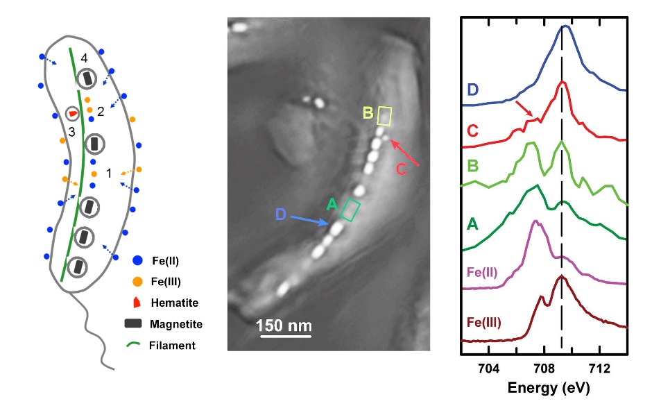
(Left) Schematic of the pathway of magnetosome biomineralization in Magnetovibrio blakemorei strain MV-1. (Center) Ptychography absorption image of a single MV-1 cell (average of 76 images from 700 to 732 eV). Four spatial regions are identified: (A) gap in the magnetosome chain, (B) precursor region, (C) immature magnetosome, and (D) mature magnetosome. (Right) Fe L3 spectra from regions A through D. [Left image courtesy Xiaohui Zhu; Middle and right images from Zhu, X., et al. “Measuring Spectroscopy and Magnetism of Extracted and Intracellular Magnetosomes Using Soft X-Ray Ptychography,” PNAS 113, E8219 (2016). DOI:10.1073/pnas.1610260114.]
The Summary
Magnetotactic bacteria (MTB) synthesize chains of magnetic nanocrystals (magnetosomes) that interact with the Earth’s magnetic field like an inner compass needle, simplifying their search for optimum environments. Some studies investigating how these magnetosomes form suggest that hematite or amorphous ferrihydrite act as precursors, while others posit that they are formed directly from solution-phase Fe(II) and Fe(III). Thus, identifying the chemical states of precursors would be a good way to differentiate among competing models.
Ptychography is a coherent diffractive imaging technique with high resolution (7 nm in this work) and excellent chemical-state sensitivity. Researchers obtained ptychographic absorption and phase spectra of magnetosomes from a marine MTB (Magnetovibrio blakemorei strain MV-1). The data, obtained from mature magnetosomes, immature magnetosomes, precursor regions, and the gaps between magnetosome chains, indicated that different iron species can coexist in a single cell. Based on the results, the researchers proposed a model in which soluble Fe(II) is taken up from the environment and partially oxidized to Fe(III), which in turn is then oxidized and transformed into hematite and ultimately into magnetite.
Researchers also used ptychography to measure the X-ray magnetic circular dichroism (XMCD) spectra of both extracellular and intracellular magnetosomes. XMCD probes the magnetism of a crystal in a site-specific manner, showing that absorption and XMCD ptychography signals provide complementary information. These experiments demonstrate that ptychography, which combines high spatial resolution, high-sensitivity chemical speciation, and site-specific magnetic information, offers a powerful probe for biomineralization studies.
Instruments and Facilities
Scanning transmission X-ray microscope (STXM) on beamline 10ID1 of Canadian Light Source at University of Saskatchewan and the Advanced Light Source Beamlines 5.3.2.1 and 11.0.2 at Lawrence Berkeley National Laboratory.
Funding
Some scanning transmission X-ray microscope (STXM) results measured at STXM on beamline 10ID1 at the Canadian Light Source (CLS), which is supported by the Canada Foundation for Innovation, Natural Sciences and Engineering Research Council (NSERC), Canadian Institute for Health Research, National Research Council, and University of Saskatchewan. Research funded by NSERC and Canada Research Chairs. Most measurements made at 5.3.2.1 and 11.0.2 beamlines at Advance Light Source (ALS), Lawrence Berkeley National Laboratory (LBNL), which is supported by the Office of Basic Energy Sciences (OBES), U.S. Department of Energy (DOE), under Contract DE-AC02-05CH11231. Work partially supported by Center for Applied Mathematics for Energy Research Applications, which is a partnership between two DOE offices: OBES and Office of Advanced Scientific Computing Research (OASCR). D.A.B. support: U.S. National Science Foundation (NSF) Grant EAR-1423939.
Related Links
References
Zhu, X., et al. “Measuring Spectroscopy and Magnetism of Extracted and Intracellular Magnetosomes Using Soft X-Ray Ptychography,” PNAS 113, E8219 (2016). [DOI:10.1073/pnas.1610260114].