X-Ray Spectroscopy Reveals Potassium Transport by Ectomycorrhizal Fungi
10/01/2024

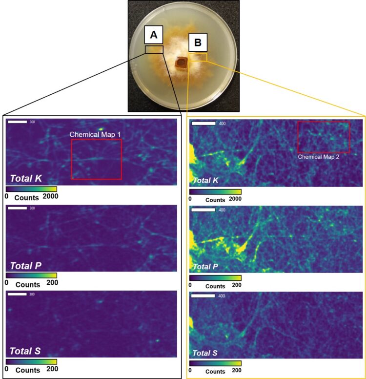
Nutrient Densities Differ with Age of Fungal Hyphae. Using X-ray fluorescence imaging, researchers produced maps of total potassium (K), phosphorus (P), and sulfur (S) in the hyphae of the fungus Paxillus ammoniavirescens. The younger, less dense hyphae (A; left panels) contained lower concentrations of K, P, and S than the older hyphae (B; right panels) and specific K chemistry differed with hyphal age as well. K and P frequently colocalized.
[Reprinted from Fungal Biology, vol. 128(6), Richardson et al., "X-ray Fluorescence and XANES Spectroscopy Revealed Diverse Potassium Chemistries and Colocalization with Phosphorus in the Ectomycorrhizal Fungus Paxillus ammoniavirescens," pp. 2054-61, Copyright 2024, with permission from Elsevier.]
The Science
Ectomycorrhizal (ECM) fungi are symbiotic microbes essential to many plants, facilitating nutrient transfer via root colonization. Critical plant nutrients, such as carbon (C), nitrogen (N), phosphorus (P), and potassium (K), are transferred through this mutualistic partnership. These nutrients also play a role in tolerance to environmental stressors and rhizosphere nutrient cycling.
Compared to N and P, the uptake, transport to plants, and storage of K by ECM is poorly studied. However, K is pivotal to several plant processes, such as enzyme activation (impacting ATP production), osmoregulation, and disease resistance. Despite this, K is limited in most environments due to its strong association with minerals. Therefore, improving and maintaining sufficient K concentrations in plants is essential for plant efficiency and vitality in a diverse range of ecosystems.
Researchers used a combination of potassium K-edge X-ray fluorescence (XRF) imaging and X-ray absorption near-edge structure (XANES) spectroscopy on P. ammoniavirescens to investigate the distribution of K chemistries across hyphal biomass. The approach revealed the presence of several K counter-ions to carboxylic acids. This may suggest that, besides their direct transfer to colonized roots, K ions can also be involved in the production of compounds necessary for sourcing nutrients from their surrounding environment by ECM fungi. Additionally, this work reveals that XANES spectroscopy can be used to identify the various forms of K accumulating in biological systems.
The Impact
This research reveals that XRF imaging and XANES spectroscopy can be used to identify the various forms of K accumulating in biological systems, something which is understudied, and shows promise in deepening understanding of the role ECM fungi play in K acquisition and transport. More established and dense ECM have several, more complicated K chemistries compared to younger individual hyphae. This suggests that older versus newer hyphae have different roles in the sourcing, transport, storage, and transfer of K between ECM and host plant symbiotic partners.
The ECM fungus Paxillus ammoniavirescens is considered a generalist that associates with many tree species, and has recently been observed to improve K nutrition and improve salinity stress in pine trees. Improving understanding of K uptake, storage, and transport mechanisms by this fungi is therefore applicable to a broad range of ECM and host plants.
The Summary
P. ammoniavirescens was grown in the absence of a plant host on a solid modified Melin-Norkans medium at 26°C for 3 weeks. Dense hyphal biomass formed on the surface of the media.
To prepare hyphal samples for synchrotron analyses, X-ray-compatible tape was placed on the dense biomass, maintaining a known spatial orientation of the mycelium. Researchers mapped the distribution of K, S, and P along hyphae and observed the accumulation of different K chemistries in different parts of the mycelium, as well as colocations of K and P.
K-nitrate (KNO3), K-C-O [such as K-tartrate K2(C4H4O6) and K-oxalate (K2C2O4)], K-S, and K-P compounds were all present in P. ammoniavirescens. In particular, K-C-O compounds were present as hotspots either on the outside of younger mycelia or within traceable hypha in the older mycelium. This is likely associated with the production and exudation of low-molecular-weight organic acids within younger mycelia, but storage and transport in older mycelia.
Funding
Use of the Stanford Synchrotron Radiation Lightsource, SLAC National Accelerator Laboratory, is supported by the U.S. Department of Energy, Office of Science, Office of Basic Energy Sciences under Contract No. DE-AC02-76SF00515. The SSRL Structural Molecular Biology Program is supported by the DOE Office of Biological and Environmental Research, and by the National Institutes of Health, National Institute of General Medical Sciences (P30GM133894). The contents of this publication are solely the responsibility of the authors and do not necessarily represent the official views of NIGMS or NIH. We also acknowledge support from the AFRI program (grants no. 2020-67013-31800 and 2022-67013-36864) from the USDA National Institute of Food and Agriculture.
Related Links
References
Richardson, J. A., Rose, B. D., and Garcia, K. 2024. “X-ray Fluorescence and XANES Spectroscopy Revealed Diverse Potassium Chemistries and Colocalization with Phosphorus in the Ectomycorrhizal Fungus Paxillus ammoniavirescens,” Fungal Biology 128(6), 2054–61. DOI:10.1016/j.funbio.2024.08.004
